
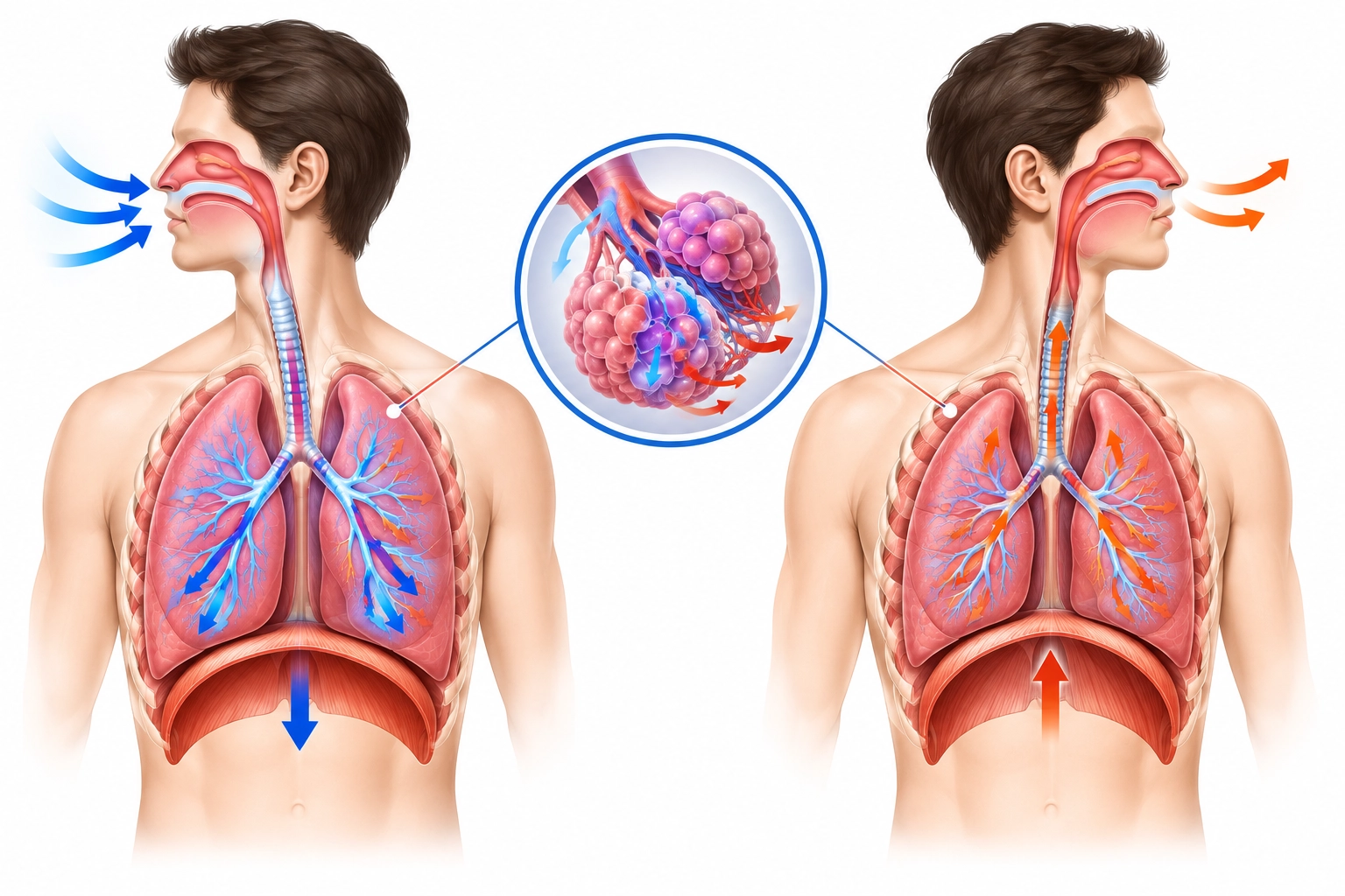
Every minute of your life, your body performs a task you rarely notice. Air flows into your chest, oxygen reaches your blood, and carbon dioxide leaves. This mechanical flow of air is called pulmonary ventilation, and it is the first step in keeping every cell alive. In this guide, you will learn how pulmonary ventilation works, which muscles drive it, and why it matters for your health. We will also explore the mechanism of breathing, the types of breathing, and the measurements doctors use.
To define pulmonary ventilation in simple words, it is the movement of air between the atmosphere and the lungs. Many people call it “breathing,” and that is a fair shortcut. Strictly speaking, pulmonary ventilation covers only the mechanical movement of air in and out. The exchange of gases across the alveolar wall is a separate step called external respiration.
Doctors measure it as minute ventilation, which equals tidal volume multiplied by respiratory rate. A healthy resting adult moves about 500 mL of air per breath and takes 12 to 16 breaths per minute. That gives roughly 6 to 8 liters of air per minute.
Define Pulmonary Ventilation in Clinical Terms
In a clinical setting, it is the total volume of air entering or leaving the lungs each minute. A fall in ventilation can signal respiratory failure, drug overdose, or nerve and muscle disease, so clinicians check it early.
Air travels through the nose, pharynx, larynx, trachea, bronchi, and bronchioles before reaching the alveoli. The lungs sit inside the thoracic cavity and are wrapped in a thin double membrane called the pleura. A film of pleural fluid keeps the lungs attached to the chest wall while letting them slide smoothly.
Muscles That Power Pulmonary Ventilation
The diaphragm is the main muscle of inspiration and does most of the work during quiet breathing. The external intercostal muscles lift the ribs. During exercise, the sternocleidomastoid and scalene muscles help. Quiet expiration is passive, but forced expiration recruits the internal intercostals and abdominal muscles.
The mechanism of breathing depends on pressure differences. Air always flows from higher pressure to lower pressure, and the body creates these gradients by changing the volume of the thoracic cavity.
Boyle’s Law and Pulmonary Ventilation
Boyle’s law states that the pressure of a gas falls when its volume rises, provided temperature stays constant. Pulmonary ventilation applies this law directly. When the chest expands, pressure inside the lungs drops below atmospheric pressure and air rushes in. When the chest shrinks, pressure rises above atmospheric and air flows out.
Mechanism of Respiration Explained
Many students confuse the mechanism of respiration with the mechanism of breathing. Respiration is the larger process. It includes ventilation, external respiration in the lungs, gas transport in blood, internal respiration at the tissues, and cellular respiration inside mitochondria. Ventilation is only the first link in that chain.
Understanding inspiration and expiration differences makes the mechanism of breathing easy to follow. The table compares both phases during quiet breathing.
Feature | Inspiration | Expiration |
Nature | Active | Passive at rest |
Diaphragm | Contracts and flattens | Relaxes and domes upward |
External intercostals | Contract | Relax |
Thoracic volume | Increases | Decreases |
Lung pressure | Below atmospheric | Above atmospheric |
Airflow | Into the lungs | Out of the lungs |
During inspiration, the diaphragm descends and the ribs swing up and out. During expiration, the muscles relax and the elastic recoil of lung tissue squeezes air out. That recoil is why quiet expiration costs almost no energy.
There are several types of breathing, and each serves a different need:
Knowing these types of breathing helps you tell when pulmonary ventilation is normal and when it is under strain.
Spirometry divides each breath into measurable parts, and these numbers show how well the lungs move air. Tidal volume is the amount of air in one quiet breath, about 500 mL. Inspiratory reserve volume is the extra air you can pull in after a normal breath, roughly 3,000 mL. Expiratory reserve volume is the extra air you can push out, around 1,100 mL. Even after a full exhale, about 1,200 mL of residual volume stays inside so the alveoli never collapse. Together, these volumes make up vital capacity, which is often about 4,600 mL in a healthy young adult and falls with age and lung disease.
You do not have to think about breathing because the brainstem handles it. The medulla oblongata contains the dorsal and ventral respiratory groups, which set the basic rhythm. The pons fine-tunes that rhythm and smooths the shift between inhaling and exhaling. Chemoreceptors in the carotid and aortic bodies, plus central chemoreceptors in the medulla, detect changes in carbon dioxide, oxygen, and pH. A rise in carbon dioxide is the strongest trigger, so you breathe faster and deeper almost at once. You can override this system briefly by holding your breath, but the urge to breathe soon takes over.
Not all inhaled air reaches the gas exchange surface. About 150 mL of every breath fills the anatomical dead space, such as the trachea and bronchi. Alveolar ventilation is the portion of pulmonary ventilation that reaches the alveoli.
The formula is (tidal volume minus dead space) multiplied by respiratory rate. With normal values, (500 − 150) × 12 equals 4,200 mL per minute. Shallow, rapid breathing is therefore inefficient. Breaths of 250 mL at 24 per minute give the same 6,000 mL minute ventilation, yet alveolar ventilation falls to only 2,400 mL.
Fresh air is useless without blood flow beside it. The ventilation perfusion ratio, written V/Q, compares alveolar ventilation with pulmonary blood flow. In a healthy lung, ventilation is about 4 L/min and blood flow about 5 L/min, giving an ideal ratio near 0.8.
Either mismatch can lower blood oxygen, so clinicians read ventilation together with perfusion.
Several factors decide how easily air moves:
Conditions such as COPD, asthma, and pulmonary fibrosis can reduce pulmonary ventilation. Spirometry is the standard test that detects these patterns.
